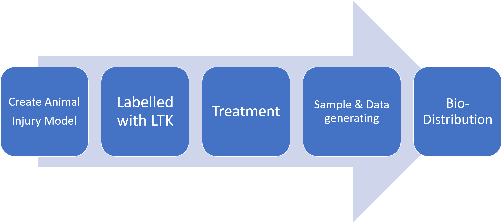
Animal injury model
Wound dressing for medical and aesthetic use
Experiment period: 2017
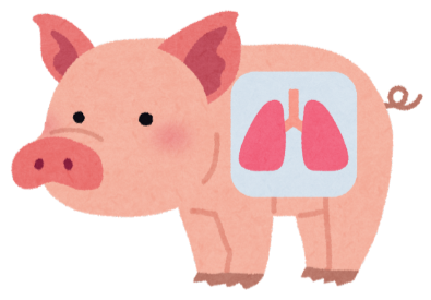
In this case, we wanted to use stem cells (MSCs) to treat damaged and hard-to-repair lung injury cells (e.g. Covid sequelae).
Previously, most scholars have used PCR to detect human DNA or immunostaining to identify human-specific nuclear proteins, but neither of these approaches can show the biodistribution of stem cells and quantitative information to assess the efficacy and safety of the drug in vivo.
When implementing large animal injury models, especially in pigs, which have many similarities to human physiology, a more accurate assay is critical, and therefore LTK was used as an important basis for tracking stem cells in this case.
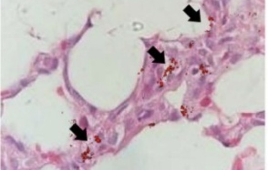
The image on the right is a composite image of a pig lung section in bright view and LTK fluorescence taken by our exclusive technique. The red dots pointed by the black arrows are the originally identified stem cells (MSCs), and the migration of stem cells was successfully observed using this technique.
Precise Labelling
The special modification on the surface of the material allows LTK to be precisely calibrated on stem cells without cytotoxicity and without cellular spitting out, making it a very stable material.
Background-free
In addition to stabilizing the fluorescence itself, the background noise can be removed with the use of exclusive technology, eliminating the need for additional post-retouching actions as with common products.
Quantitative analysis
In addition to clearly showing the distribution of cells, we can also collect samples from various organs for quantitative services to understand the migration of cells throughout the organism.


 02-26558597
 115 F17-1, No.3, Park St.Nangang Dist., Taipei, Taiwan